Welcome to the Secretory Function module. Today we travel the length of the GI tract — from saliva in the mouth, to acid and enzymes in the stomach, to bile and pancreatic juice in the intestine — and see how each secretion is produced, regulated, and why it matters. Every scene maps to one of the objectives below.
Lecture Objectives
Explain how salivary secretions are produced, modified, and regulated (ANS input, ductal cell function).
Identify the major secretory cells, their products, and how secretion changes across the cephalic, gastric, and intestinal phases.
Describe the roles of digestive enzymes and bicarbonate, and how CCK and secretin regulate their release.
Explain how bile is formed, stored, and released, and why it's critical for fat digestion and waste excretion.
Differentiate secretions of the small vs. large intestine, including protective mucus and fluid/electrolyte balance.
Apply secretory physiology to disorders such as (but not limited to) cystic fibrosis, gallstones, and secretory diarrhea.
📖 Supplemental Readings
Boron & Boulpaep, Medical Physiology, 3rd ed. — Ch 43: Pancreatic & Salivary Glands. Recommended for especially clear coverage of secretory physiology.
Guyton & Hall, Medical Physiology, 14th ed. — Ch 65: Secretory Functions of the Alimentary Tract.
Costanzo Physiology, 8th ed. — Ch 8: Gastrointestinal Physiology. Recommended as a concise, exam-focused review.
You'll move through 20 scenes with interactive figures, predict-and-reveal questions, a recall check, and a clinical integration case. Budget about 55–60 minutes if you use the narration and AI-guided feedback. Your progress saves automatically, so you can close the tab and return anytime.
🎧 Highly Recommended
Use the Listen button on teaching scenes that include narration, much like having the instructor walk you through the material. Recall, clinical, and summary scenes may focus on active work instead of narration.
Privacy: This module does not send navigation, answer, score, or AI conversation data to its feedback system. If you choose to submit the optional end-of-module feedback, your rating and written comment are sent to a restricted-access Google Sheet with a randomly generated attempt identifier. Your name and student ID are not requested. Do not enter names, student IDs, patient information, or other identifying or sensitive information in feedback or AI fields.
Before we dive into each gland, let's zoom out. A handful of control principles apply everywhere in the GI tract — from saliva in the mouth, to gastric juice in the stomach, to pancreatic secretions in the intestine.
Sensory and local stimulation. In the mouth, food activates taste and touch receptors. That sensory input reaches the brainstem salivatory nucleiBrainstem centers that integrate oral sensory input and increase autonomic output through CN VII and IX to the major salivary glands., which increase autonomic output to the major salivary glands. Farther down the GI tract, luminal contact also activates local epithelial and enteric signals.
Enteric reflexes. In the stomach and small intestine, the enteric nervous systemThe gut's own "little brain" — neural networks in the wall that sense stretch and chemical signals and fine-tune secretion locally. plays a bigger role, responding to stretch and chemical signals.
Autonomic control. Parasympathetic pathways generally increase GI secretion. The vagus supplies much of the stomach and proximal intestine, while CN VII and IX control the major salivary glands and pelvic parasympathetic pathways influence the distal colon. Sympathetic activity generally reduces net GI secretion, especially through vasoconstriction. The salivary glands are a notable exception: sympathetic input can directly stimulate a smaller, protein-rich secretion even though intense vasoconstriction may reduce overall flow.
Hormonal regulation. In the mouth, hormones matter little. But downstream, GI hormones like gastrin, secretin, and CCK powerfully amplify or inhibit secretion depending on what's in the meal.
Now let's look at the basic cellular mechanism of secretion. This should feel familiar from biochemistry — we're just applying protein synthesis and secretion to the GI system.
It starts inside the glandular cell, which — like every epithelial cell we'll meet today — has two distinct sides. The basolateralThe side of the cell facing the blood supply and underlying tissue — where nutrients, hormones, and nerve signals arrive, and where the Na⁺/K⁺-ATPase usually sits. side faces the blood and the underlying tissue; the apicalThe side of the cell facing the lumen — the free surface where the finished secretory product is actually released. You'll see "apical" and "basolateral" constantly for the rest of this module. side faces the lumen, where the secreted product ends up. Nutrients, powered by ATP from abundant mitochondria, fuel synthesis of proteins and enzymes in the rough ER. These products are packaged and modified in the Golgi apparatus and stored in vesicles at the cell's apical end.
The vesicles wait in storage until the cell is stimulated — by a nerve signal or a hormone. That stimulation raises intracellular calciumCa²⁺ is the universal trigger: its rise allows the storage vesicle to fuse with the apical membrane and release contents into the lumen., and calcium is the trigger that allows the vesicle to fuse with the cell membrane and release its contents into the lumen by exocytosis.
That describes protein and mucus secretion, but it is only one of the two cellular routes we will use in this module. Glandular cells also move electrolytes through membrane pumps, channels, and exchangers; water may then follow the resulting osmotic gradient. Enzymes and mucins therefore leave in vesicles, while H⁺, HCO₃⁻, Cl⁻, Na⁺, and fluid are handled through membrane transport. Both routes require a polarized cell with distinct basolateral and apical sides.
Route 1: Protein or mucus cargo
Route 2: Electrolyte and fluid transport
Vesicles release macromolecules; membrane transport builds electrolyte-rich secretions. Many glands use both.
Every gland we'll cover in this module is built from the same handful of cell types, so let's define them here first. Acinar cells are the ones that actually make the primary secretion, and in the salivary glands they come in two flavors. Serous cellsWatery enzyme juice — thin fluid rich in α-amylase, which begins starch digestion. secrete a thin, watery fluid rich in α-amylase (starch digestion) — think "watery enzyme juice." Mucous cellsSlippery protection — thick, viscous secretion that coats and lubricates food. secrete a thick, viscous fluid that coats and lubricates — think "slippery protection." Most glands contain a mix of both cell types side by side.
The three major gland pairs make about 90% of saliva. The parotidLargest salivary gland; almost entirely serous acinar cells — the main source of α-amylase. is almost entirely serous acinar cells — the main source of α-amylase. The submandibular gland is a mix of serous and mucous acinar cells. The sublingual gland is also mixed, but its acinar cells skew more mucous-heavy. On average, together they make about 1 liter of saliva per day.
Ductal cells then modify that primary saliva as it flows past them: they reabsorb Na⁺ and Cl⁻ and secrete K⁺ and HCO₃⁻. Myoepithelial cells wrap around the acini and ducts and contract to squeeze saliva forward into the mouth.
So saliva isn't just water — it's a balanced mix of watery enzyme-rich fluid, protective mucus, and electrolyte adjustments. Its jobs include lubrication, pH buffering, early starch digestion through α-amylase, antimicrobial defense, tooth protection, and taste. Lingual lipase from the lingual glands also enters the oral cavity and begins fat digestion, but it is not a major-gland acinar product.
Where are the major salivary glands?
Three paired glands produce most saliva. Their locations differ, as does the balance of watery serous secretion and lubricating mucous secretion.
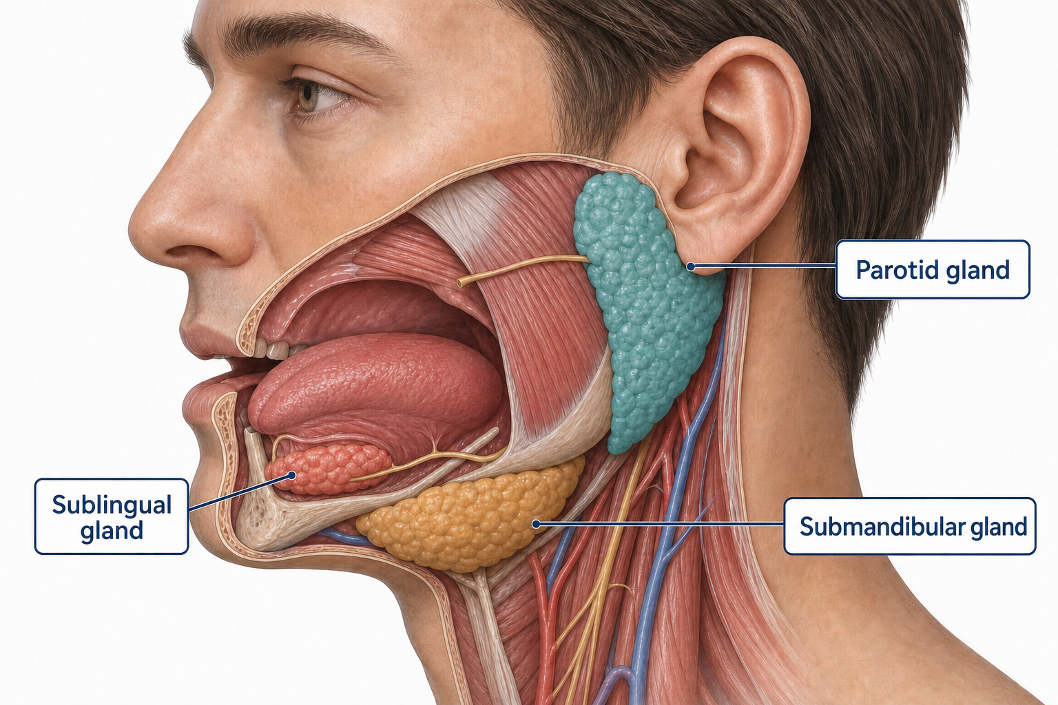
Parotid gland
Location: Near the ear, with its duct crossing the cheek toward the mouth.
Composition: Predominantly serous—watery and enzyme-rich; a major source of α-amylase.
Location changes and the serous–mucous balance changes, but all three glands use the same basic secretory architecture.
What is inside a salivary gland?
Zoom in and the shared plan becomes clear: acinar cells make primary saliva, myoepithelial cells help propel it, and a branching duct system carries it toward the mouth.
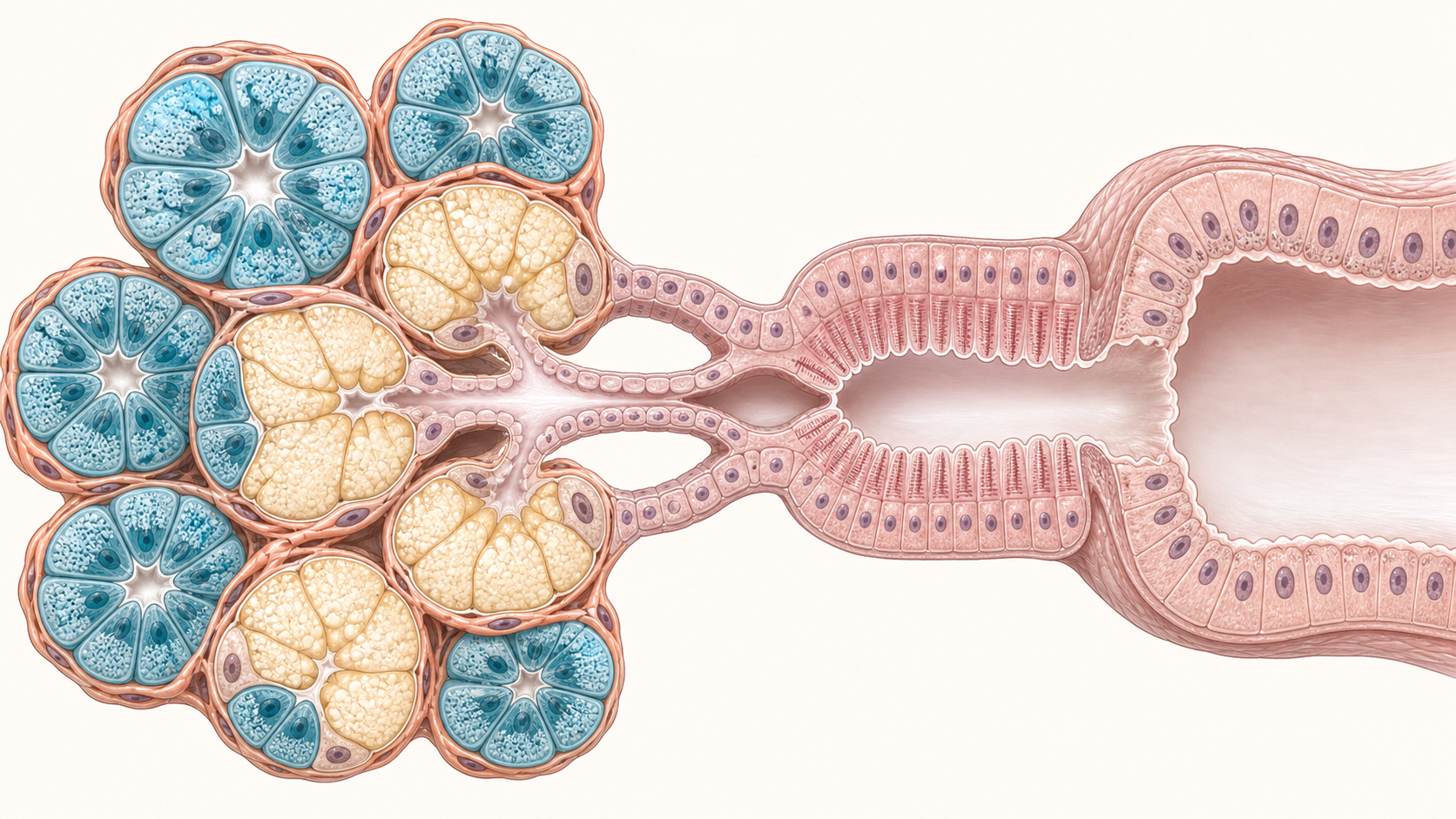
Serous acinar cells
Where: Wedge-shaped secretory cells surrounding the acinar lumen.
Job: Produce a thin, watery secretion rich in enzymes such as α-amylase.
Acinar cells create primary saliva, myoepithelial cells help move it forward, and duct cells modify it on the way to the mouth.
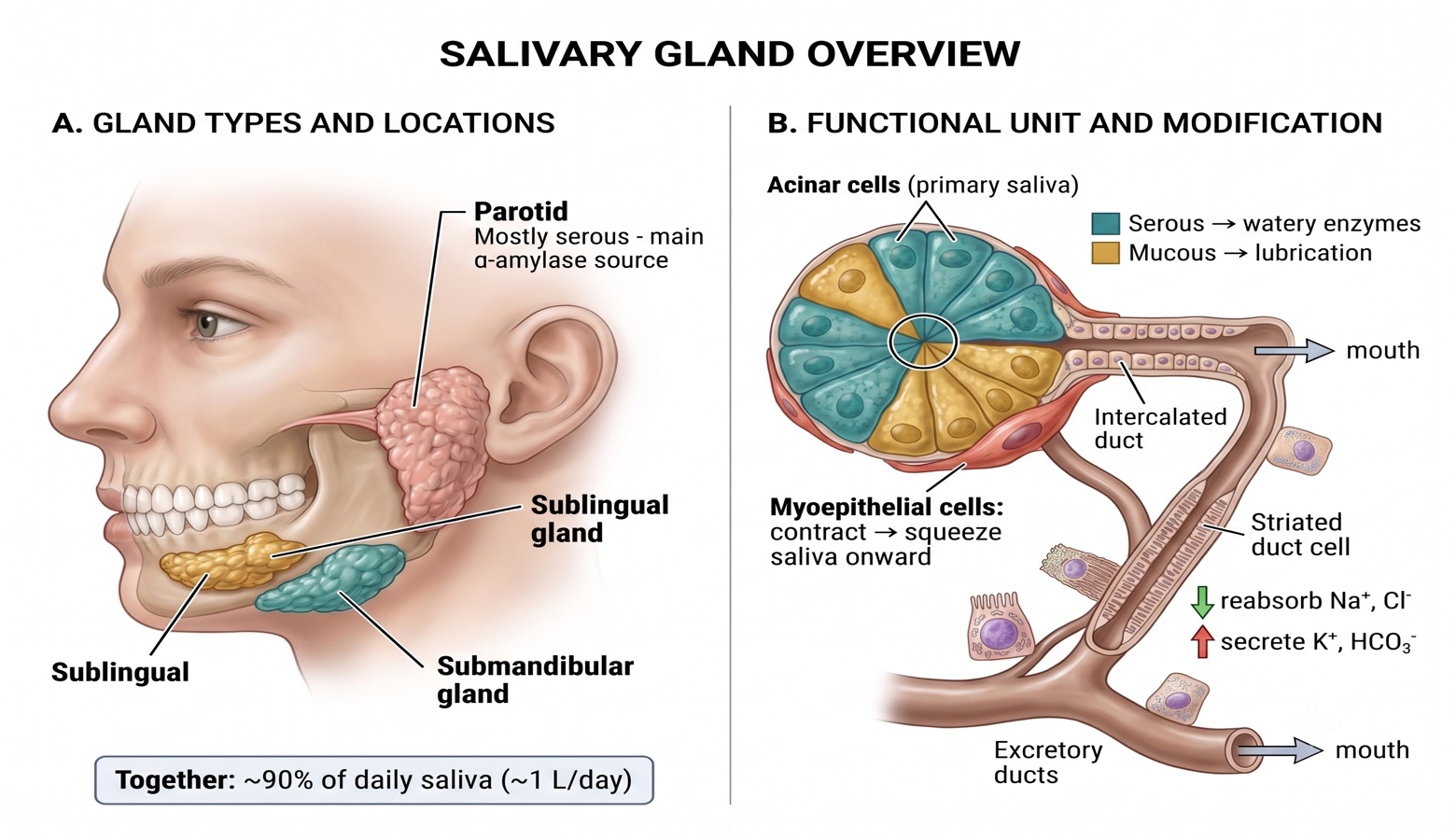
Saliva is made in two steps. First, the acini — grape-like clusters of secretory cells — produce the primary salivaIsotonic, plasma-like fluid plus major-gland organic components such as α-amylase, mucins, IgA, lactoferrin, and kallikrein.. This primary fluid is isotonic, with an electrolyte composition close to plasma, and it carries major-gland organic components including amylase, mucins, and antimicrobial factors such as IgA and lactoferrin.
Second, the fluid flows into the ductal cells, which fine-tune it. On the basolateral side, the Na⁺/K⁺ ATPase maintains the gradients that drive everything. On the apical side, the net effect is: Na⁺ and Cl⁻ are reabsorbed, while K⁺ and HCO₃⁻ are secreted.
Here's the critical point: the tight junctions of these duct cells are impermeable to waterIons move, but water cannot follow — so the fluid becomes progressively more dilute (hypotonic) as it travels down the duct.. So even though ions move, water can't follow — and the final saliva that reaches your mouth is hypotonic compared to plasma.
Isotonic primary saliva
Its electrolyte concentration initially resembles plasma. Acinar cells also add the molecules that begin digestion and protect the oral cavity.
Reabsorbed from the lumen toward the interstitium.
Blocked. Water-impermeable duct epithelium prevents water from following the ions.
Secreted from the duct cells into the lumen.
Step 1: the acinus produces isotonic primary saliva and supplies digestive and antimicrobial components.
The duct removes more NaCl than it adds other ions, but relatively little water follows. That combination, net salt reabsorption plus low ductal water permeability, is what makes final saliva hypotonic.
Here's a high-yield concept: the composition of saliva changes with flow rate. Salivary glands are remarkably active — up to 1 mL of saliva per gram of tissue per minute — and sustaining that requires increased blood flow, driven by parasympathetic stimulation.
At low flow, saliva lingers in the ducts, giving ductal cells lots of time to reabsorb Na⁺ and Cl⁻ and secrete K⁺. The result is very hypotonic saliva that looks quite different from plasma. At high flow, saliva rushes through with little time for modification, so it's less hypotonic and more plasma-like. The one exception is bicarbonate, which actually rises at high flow — protective, since it helps neutralize acid.
Drag the flow-rate slider below and watch the four ions change.
At low flow, prolonged duct contact produces extensive ion modification and very hypotonic saliva.
Curves show approximate teaching relationships rather than fixed laboratory reference values. Dashed lines indicate each ion's plasma concentration.
Explain the pattern before choosing
Why does higher salivary flow make Na⁺ and Cl⁻ concentrations more plasma-like?
Salivary secretion is regulated almost entirely by the autonomic nervous system — and here's the twist. In most organs, sympathetic and parasympathetic oppose each other. In the salivary glands, both actually stimulate secretion — but the parasympathetic system is by far the stronger driver.
Parasympathetic input travels through cranial nerves VII (facial) and IX (glossopharyngeal), releasing acetylcholine. It produces a large volume of watery saliva and boosts blood flow to the glands. This is the cephalic phaseSalivation triggered by the sight, smell, taste, or even the thought of food — before anything reaches the stomach. response — smelling, tasting, or chewing food.
The sympathetic system, via the superior cervical ganglion and norepinephrine, also stimulates secretion through adrenergic receptors. β-adrenergic signaling particularly promotes protein release, while α-adrenergic vasoconstriction can limit glandular blood flow. The result is usually a smaller volume of thicker, more viscous saliva, so sympathetic activity changes texture more than volume.
Salivation is inhibited by dehydration, sleep, fear, and anticholinergic drugs like atropine.
Because salivation is parasympathetic-driven, blocking it causes dry mouth — which is why patients on anticholinergics, many antihistamines, and some antidepressants complain of it. Autoimmune destruction of the glands in Sjögren's syndrome causes profound, chronic xerostomia.
The stomach has four jobs that matter for this block: it begins protein digestion (pepsin), it creates a strongly acidic environment that kills swallowed microbes and activates pepsinogen, it secretes intrinsic factor — the one gastric product with no backup elsewhere in the body, required for B₁₂ absorption — and it protects its own lining with a mucus/bicarbonate barrier.
Each job belongs to a different cell type living in the gastric pits and glands:
Mucous cells (surface mucous and mucous neck cells) secrete mucus and HCO₃⁻Together they form a protective gel layer that buffers the epithelium against the highly acidic lumen. — the stomach's shield against its own acid. Parietal cells (also called oxyntic cells) secrete HCl and intrinsic factor. Chief cells secrete pepsinogen, the inactive precursor of pepsin, plus a small amount of gastric lipase. G cells secrete gastrin — not into the lumen, but into the blood, where it circulates back to stimulate parietal cells.
Two more cells act as local paracrine controllers of acid. ECL cellsEnterochromaffin-like cells — paracrine cells nestled near parietal cells that release histamine, the most powerful amplifier of acid secretion. We'll return to them in the next scene on the parietal cell. sit beside the parietal cells and release histamine — the single biggest amplifier of acid secretion, and the key to the next scene. D cellsDelta cells scattered through the gastric glands. Somatostatin is the universal inhibitor — a useful memory hook is "D = Don't secrete." release somatostatin, the universal "off switch": when the lumen gets too acidic, somatostatin inhibits gastrin, histamine, and acid — a built-in negative-feedback brake.
These cells aren't spread evenly. The acid machinery — parietal, chief, and ECL cells — clusters in the oxyntic glandsThe acid-secreting glands of the fundus and body. They contain parietal, chief, ECL, D, and mucous neck cells. of the fundus and body. G cells are concentrated in the antrum. D cells occur in both regions: antral D cells inhibit gastrin release, while oxyntic D cells provide a local somatostatin brake on ECL and parietal-cell signaling. Here's the map:
Match each gastric cell type to its secretory product.
Drag the product on the right and drop it onto the correct cell type on the left. Tap on mobile: tap a product, then tap a slot.
Cell Type
Secretory Product
Gastric secretion follows the meal. The brain primes the stomach, food in the stomach produces the strongest response, and the duodenum eventually applies the brake. Use the timeline to follow where each signal begins and what it does.
Read the full explanation
Gastric secretion unfolds in three overlapping phases, named for where the triggering stimulus originates.
The cephalic phase (~30% of the response) fires before food ever reaches the stomach — the sight, smell, taste, or even thought of food, plus chewing and swallowing, drive the vagus nerve to directly stimulate parietal cells (ACh) and G cells (via GRP/bombesin) to release gastrin. It's a purely neural, feedforward signal that primes the stomach in advance.
The gastric phase (~60%, the dominant phase) begins once food is actually in the stomach. Distension activates stretch receptors, driving vagovagal and local enteric reflexes, while digested protein fragments directly stimulate G cells to release even more gastrin. Most of the meal's total acid output happens here.
The intestinal phase (~10%) is smaller and biphasic. Early on, protein products entering the duodenum weakly stimulate a little more gastrin release. But as chyme accumulates — especially if it's acidic, fatty, hypertonic, or distending the duodenum — the enterogastric reflexA neural reflex, plus hormones like secretin, CCK, and GIP, that together inhibit further gastric secretion and slow gastric emptying once the duodenum is loaded with chyme. and hormones like secretin, CCK, and GIP kick in and inhibit further gastric secretion and emptying — protecting the duodenum from being overwhelmed.
Cephalic
Gastric
Intestinal
Anticipate → respond strongly → prevent the duodenum from being overwhelmed.
Surgical vagotomy was once a mainstay treatment for peptic ulcer disease — cutting the vagus removes the cephalic phase and much of the vagal drive in the gastric phase, sharply reducing acid output. It's now largely obsolete, replaced by PPIs, H₂ blockers, and H. pylori eradication.
The parietal cell is a single-cell acid factory. It pumps H⁺ into the gastric lumen against one of the steepest ion gradients in the body, producing gastric juice as concentrated as ~150 mM HCl — a pH near 0.8.
Here's the mechanism. Inside the cell, CO₂ + H₂O combine (catalyzed by carbonic anhydraseA very fast enzyme that rapidly interconverts CO₂ + H₂O and H₂CO₃, supplying H⁺ and HCO₃⁻. It is not the regulated final step in acid secretion.) to form H₂CO₃, which dissociates into H⁺ and HCO₃⁻. The H⁺ is then actively pumped into the lumen by the apical H⁺/K⁺-ATPase — the regulated final common pathway — in exchange for K⁺, which recycles back out through apical K⁺ channels. Cl⁻ follows H⁺ into the lumen through apical Cl⁻ channels, and the two combine to form HCl.
Meanwhile, the HCO₃⁻ generated inside the cell is shuttled out across the basolateral membrane by a Cl⁻/HCO₃⁻ exchanger — pulling Cl⁻ in to resupply the apical side, and releasing HCO₃⁻ into the venous blood. This is the alkaline tideThe transient rise in blood/venous pH after a meal, caused by HCO₃⁻ leaving parietal cells into the bloodstream as acid is secreted into the lumen. — the brief rise in blood pH you'd measure after a big meal.
Three signals converge on this pump: acetylcholine (vagus, M₃ receptors, ↑Ca²⁺), gastrin (blood-borne, CCK₂ receptors, ↑Ca²⁺), and histamine (paracrine, from neighboring ECL cells, H₂ receptors, ↑cAMP). Histamine is the biggest amplifier — it potentiates the other two, which is exactly why H₂ blockers are so effective and why gastrin or ACh alone produce only a modest response without it.
Use the simulator below: stimulate normal acid secretion, then try blocking the pump the way a PPI would.
PPIs (e.g., omeprazole) irreversibly bind the H⁺/K⁺-ATPase itself — the final common pathway — so they block acid secretion no matter which of the three stimuli is driving it. H₂ blockers only remove the histamine input, which is why they're effective but generally less potent than PPIs.
Identify the final common pathway
ACh, gastrin, and histamine use different receptors. Why can blocking the H⁺/K⁺-ATPase still shut down the acid response to all three?
Chief cells (also called peptic cells) sit deep in the gastric glands and synthesize and secrete pepsinogenAn inactive zymogen — it must be cleaved before it can digest protein, which prevents chief cells from digesting themselves., an inactive precursor, along with a small amount of gastric lipase.
Pepsinogen only becomes active pepsin when it meets the acidic environment (pH < 5, optimal around 2–3) created by the neighboring parietal cells. Once a little pepsin has formed, it can cleave more pepsinogen itself — an autocatalytic, positive-feedback activation that rapidly amplifies the supply.
Pepsin is an endopeptidase: it cleaves proteins into smaller peptides, not free amino acids. Its window of action is narrow because substantial activity requires an acidic environment. Once pancreatic bicarbonate raises duodenal pH, pepsin activity falls while pancreatic proteases continue protein digestion.
Chief-cell pepsinogen secretion is stimulated most directly by ACh from vagal and enteric cholinergic pathways. Gastrin and secretin can also support pepsinogen secretion. Luminal acid promotes gastric reflex signaling and, separately, provides the low pH that converts released pepsinogen into active pepsin. Because cholinergic activity drives both parietal and chief cells, pepsinogen release is coordinated with the acid needed to activate it.
Before we move on to the pancreas and bile, let's lock in Part 3. Try these five questions from memory before reviewing earlier scenes.
The exocrine pancreas is built from the same two-cell-type logic you've already seen twice — first in the salivary glands, then in the stomach. Here it's acinar cells and duct cells, and together they make up to 1.5 L/day of pancreatic juice.
Acinar cells synthesize and secrete digestive enzymes for proteins, carbohydrates, fats, and nucleic acids. The major groups in this lecture are proteases, amylase, lipase, and nucleases.
Duct cells have the other job: they secrete a fluid rich in HCO₃⁻ that neutralizes the acidic chyme arriving from the stomach — protecting the duodenal mucosa and raising the pH into the range these enzymes actually need to work. We'll look at exactly how that's regulated in the next scene.
Pancreatic duct cells create an alkaline, bicarbonate-rich fluid that neutralizes gastric acid in the duodenum. Carbonic anhydrase generates H⁺ and HCO₃⁻ inside the cell, while additional HCO₃⁻ can enter from the blood.
At the apical membrane, a Cl⁻/HCO₃⁻ exchanger moves bicarbonate into the duct lumen. CFTR supports chloride recycling, keeping luminal Cl⁻ available for that exchange. Sodium and water follow, producing an isosmotic alkaline secretion.
Use the comparison below to follow the first transport change before predicting the downstream fluid response.
Start at the apical membrane: CFTR keeps chloride available in the lumen, supporting the exchanger that secretes bicarbonate. Sodium and water then follow.
Predict the consequence
If CFTR support for chloride recycling is reduced, explain how pancreatic duct secretion changes.
Read the full explanation
What turns this on? When acidic chyme (pH < 4.5) hits the duodenum, S cells in the duodenal mucosa release secretin into the blood. Secretin acts on duct cells (via cAMP) to drive a large-volume, HCO₃⁻-rich, enzyme-poor secretion — neutralizing the acid and protecting the mucosa.
Separately, when fatty acids and amino acids from a meal hit the duodenum, I cells release CCK (cholecystokinin). CCK's main pancreatic target is the acinar cell — it drives a low-volume, enzyme-rich secretion. CCK also contracts the gallbladder and relaxes the sphincter of Oddi, which we'll cover next scene.
The two hormones don't just act in parallel — they potentiate each other: CCK amplifies the ductal response to secretin, and secretin amplifies the acinar response to CCK. Between meals, low-level vagal (ACh) tone keeps some baseline secretion going too.
Run the meal through the duodenum
Choose the luminal stimulus, predict the dominant pathway, and then reveal the coordinated pancreatic response.
| Hormone | Released by | Trigger | Main Pancreatic Target | Effect |
|---|---|---|---|---|
| Secretin | S cells (duodenum) | Duodenal acid (pH < 4.5) | Duct cells | ↑ Volume, ↑ HCO₃⁻, watery, low enzyme |
| CCK | I cells (duodenum) | Fatty acids & amino acids | Acinar cells | ↑ Enzymes, low volume; also contracts gallbladder |
Explain mixed-meal potentiation
An acidic meal containing fat and protein activates secretin and CCK together. Why is the pancreatic response more than simple addition while each hormone still retains its predominant target?
Hepatocytes continuously secrete roughly 600–1,000 mL of bile per day into tiny channels called bile canaliculi. Bile is a mix of bile acids/salts (made from cholesterol), bilirubin (a heme breakdown product — bile's main pigment), cholesterol, phospholipids, and IgA, plus an HCO₃⁻-rich fluid added by bile duct cells (again, secretin-responsive — the same logic as the pancreatic ducts).
Between meals, the sphincter of Oddi is closed, so bile backs up and diverts into the gallbladder, which stores it and — critically — concentrates it up to 10–20 fold by reabsorbing water and electrolytes across its epithelium.
When fat and protein hit the duodenum, CCK — the same hormone driving pancreatic enzyme release — does double duty here: it triggers gallbladder contraction and relaxation of the sphincter of Oddi, delivering a concentrated bolus of bile into the duodenum right when it's needed.
Bile's essential job is emulsification: bile salts are amphipathic, breaking large fat globules into smaller droplets to increase the surface area available for pancreatic lipase. Bile also supports absorption of fatty acids, monoglycerides, cholesterol, and fat-soluble vitamins, and provides an excretion route for cholesterol and bilirubin.
Follow one unit of bile
First, follow stations 1 → 2 → 3 → 4 in order. After reaching the duodenum, tap any station to review the pathway.
Continuous formation
Hepatocytes secrete bile acids, phospholipids, cholesterol, bilirubin, and other solutes into canaliculi. Production continues between and after meals.
State: bile is being formedCholesterol imbalance relative to bile salts can allow cholesterol to precipitate and form gallstones. The location of obstruction determines whether bile flow alone or both bile and pancreatic drainage are affected.
Brunner's glands are submucosal glands found only in the duodenum — right where acidic gastric chyme first arrives. They secrete an alkaline, HCO₃⁻-rich mucus that neutralizes acid and protects the duodenal mucosa, working alongside pancreatic HCO₃⁻. They're stimulated by local irritation, parasympathetic (vagal) input, and secretin, but inhibited by sympathetic activity.
The crypts of Lieberkühn lie at the base of villi throughout the small intestine. Goblet cells provide mucus, while crypt enterocytes secrete water and electrolytes. Enterocytes on the villi rapidly reabsorb much of that fluid while absorbing nutrients.
Crypt cells also secrete an isotonic, Cl⁻-driven fluid (via CFTR, the same channel from the pancreatic and salivary ducts) that provides the liquid medium enzymes and nutrients need — normally dwarfed by the much larger absorptive capacity of the villi, so the small intestine is a net absorber overall.
Cholera toxin locks the stimulatory G protein Gsα in its active state. Persistent adenylate-cyclase activity raises cAMP, activates CFTR, and drives massive Cl⁻ and fluid hypersecretion — the mechanism behind cholera's profuse secretory diarrhea. Absorption in the villi can't keep up with crypt output turned up this high.
By the time chyme reaches the colon, the job has changed: this is no longer about digestion — the enzymes and hormones from earlier scenes have already done their work. The large intestine's secretory role narrows to lubrication and protection.
Colonic crypts contain abundant goblet cells, which produce mucus to ease passage of fecal material, protect against mechanical damage, and help shield the mucosa from the dense bacterial environment. The mucus contains some HCO₃⁻, which helps buffer acids produced by bacterial activity.
Secretion increases with local mechanical/tactile stimulation, enteric reflexes, and parasympathetic input through the pelvic nerves. Irritation can also increase water and electrolyte secretion along with mucus, contributing to diarrhea.
Enteritis or other mucosal irritation can increase mucus, water, and electrolyte secretion. The resulting diarrhea reflects an exaggerated secretory response rather than digestive-enzyme secretion by the colon.
Use the transport principles from the salivary and pancreatic sections to follow one channel defect across multiple secretory tissues.
A person with cystic fibrosis has reduced CFTR function in pancreatic duct cells and develops thick pancreatic secretions, reduced bicarbonate delivery, and fat malabsorption with steatorrhea.
You've completed the entire module — from saliva in the mouth to mucus in the colon. Here's your performance, plus a chance to check your own confidence on each major topic.
Module Complete
Your first-attempt results and review links are ready below. You can revisit any scene without changing your original score.
Where to Focus Your Review
Module Concept Map
| System | Key Cells / Structures | Primary Regulator | Clinical Correlate |
|---|---|---|---|
| Salivary Glands | Acinar (serous/mucous), ductal, myoepithelial | Parasympathetic (CN VII, IX) | Xerostomia (Sjögren's, anticholinergics) |
| Stomach | Parietal, chief, mucous, G, D, ECL cells | ACh / gastrin / histamine; somatostatin brake | PPIs & H₂ blockers; autoimmune parietal-cell loss → intrinsic-factor loss and vitamin B₁₂ deficiency |
| Exocrine Pancreas | Acinar (enzymes), duct (HCO₃⁻) | CCK (acinar) / secretin (duct) | CF exocrine insufficiency |
| Biliary System | Hepatocytes, gallbladder | CCK (contraction) | Cholesterol gallstones |
| Small Intestine | Brunner's glands, crypt enterocytes, villus enterocytes, goblet cells | Vagal / secretin / local reflexes | Cholera — secretory diarrhea |
| Large Intestine | Goblet cells and crypts | Local mechanical stimulation / pelvic parasympathetic input | Enteritis or irritation — increased secretion |
Confidence Self-Check
Rate your confidence 1–5 on each objective. Be honest — this is for you, not for a grade.
Help Improve This Module
This feedback is optional and does not affect your course grade. Your name and student ID are not requested. Responses are associated only with a randomly generated attempt identifier and will be reviewed in aggregate to improve the module. Do not include names, student IDs, patient information, or other identifying or sensitive information.